The contents of this article are informational only and are not intended to be a substitute for professional medical advice, diagnosis, or treatment recommendations. This editorial presents the views and experiences of the author and does not reflect the opinions or recommendations of the publisher of Ophthalmology 360.
By Steven Ferrucci, OD, FAAO
Age-related macular degeneration (AMD) is a leading cause of irreversible blindness among older adults, and as we lead longer lives, the prevalence of the disease is likely to increase. Globally, it has been estimated that the number of individuals with AMD will grow approximately 50% to 288 million between 2020 and 2040.1
In America, just under 20 million people are estimated to have AMD.2 For approximately 1.2 million of these individuals, the disease has progressed to include geographic atrophy (GA).3
Given the size of the affected populations, we must ensure that AMD receives the same attention as other eye diseases such as glaucoma and diabetic retinopathy. Alongside diligent diagnosis and the consideration of both new and established pharmaceuticals, it is important to educate patients about holistic treatments. Nutraceuticals containing antioxidant vitamins, minerals, and carotenoids that have been shown in clinical trials to slow the progression of AMD are an important part of holistic treatment.
Diagnosing AMD
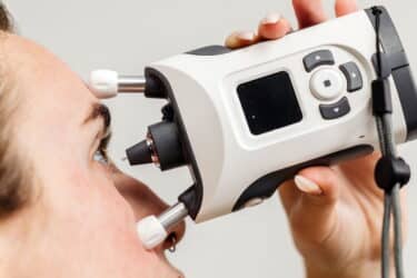
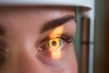
Diagnosis of both neovascular, or wet, and non-vascular, or dry, forms of AMD involve clinical examination, often with the help of advanced imaging techniques. It is standard practice to perform a dilated fundus examination to look for drusen or changes to the retinal pigmented epithelium (RPE).
Dark adaptation testing is also helpful, as impaired dark adaptation is one of the earliest signs of AMD. This testing can identify functional changes in the retina even before structural changes such as drusen become visible upon exam. Additionally, I like to get a cross-sectional view of the retina with optical coherence tomography (OCT), as it allows me to follow drusen for progression as well as look for any fluid, suggestive of conversion to wet AMD.
After diagnosing AMD, we must monitor patients to catch the transition from dry to wet AMD. This neovascular form only affects 10% to 15% percent of patients with AMD,4 but it is responsible for nearly 90% of severe damage to central visual acuity.5 The Amsler Grid is commonly used to detect early distortions of central vision, which can indicate progression from dry to wet AMD, and new products, such as the ForeSee Home by Notal Vision, are available that use other measurements to identify this conversion.
Genetic tests such as those from ArcticDx and Visible Genomics are an option to evaluate a patient’s risk of progression to advanced disease. The results can be used to set exam frequency for a patient. I am especially likely to use these tests on patients with a family history of AMD.
Treating the Disease
Wet AMD has a well-established standard of care, which is serial anti-vascular endothelial growth factor (VEGF) injections. Several newer treatments that offer longer durability to decrease patient burden are currently being investigated.
Until recently, no pharmaceutical treatments were available for dry AMD. In 2023, 2 therapies for GA received FDA approval. SYFOVRE® (pegcetacoplan; Apellis Pharmaceuticals) and IZERVAY™ (avacincaptad pegol;Astellas) have both been shown to slow the progression of GA, although they do not restore lost vision.6,7
Treatments for other forms of dry AMD involve lifestyle modification and supplementation. Smoking is perhaps the most important modifiable risk factor. Smokers can be up to 4 times more likely to develop AMD, and the risk increases with the number of cigarettes smoked each day.8 Smoking has been shown to not only increase the risk of the onset of AMD but also the likelihood of progression to advanced stages such as GA and wet AMD.9 Risk can be reduced when a patient stops smoking, although former smokers do remain at higher risk than those who have never smoked.10
AMD can also be treated with changes to diet and exercise that often serve to both prevent the onset and slow the progression of the disease. Those who eat more saturated fats and cholesterol, as well as those with a higher body mass index, have been shown to face an increased risk of AMD.11 I encourage my patients to eat green leafy vegetables, limit the amount of processed foods they eat, and decrease the amount of red meat they eat, while increasing their intake of fresh fish. I also encourage them to be physically active. Individuals who exercise at least 3 times a week are less likely to develop wet AMD than those without an active lifestyle.12
The Importance of Supplementation
In 2001, the Age-Related Eye Disease Study (AREDS), a long-term clinical trial, reported that high-risk patients taking antioxidant and zinc supplements improved their chances of retaining macular health by 25% and preserving vision by 19%.13 The second AREDS trial (AREDS2) looked at whether adding lutein and zeaxanthin to the original AREDS formula could reduce the risk of progression to AMD by more than the original 25%. Analysis showed that lutein and zeaxanthin offer protective benefits and were the only added nutrients to exceed this 25% threshold.14
Supplementation with the original AREDS formula, altered to include lutein and zeaxanthin in place of beta-carotene, is recommended by the National Eye Institute as the standard of care for individuals with at least intermediate AMD.
I often start patients on supplementation before they reach the intermediate stage. I prefer to be more proactive in these patients and start a little earlier, rather than simply watching until it progresses to a more advanced level. An AREDS2-based supplement with lutein and zeaxanthin such as MacularProtect (ScienceBased Health) is a great choice. Since many of my patients are already taking a daily multivitamin, it is easy and logical to recommend a product that combines a multivitamin with AREDS2 supplementation, such as MacularProtect Complete (ScienceBased Health).
In the past, I have not aggressively included AREDS supplements in treatment plans for those with GA, because initial analysis of the trials did not show the supplements to be of benefit to these patients. However, a recent post-hoc analysis of the AREDS trials has now reported that AREDS supplements slowed the progression of GA toward the fovea by approximately 55% over 3 years in dry AMD patients with extra-foveal lesions.15
Slowing the progression of lesions to the fovea should allow us to stave off poor vision and allow these patients more time with good, functional vision. I now continue supplementation with these nutraceuticals in patients with late-stage AMD, especially with patients whose GA is extra-foveal.
Encouraging Nutraceutical Compliance
I have found compliance to be good with nutraceuticals, as many of my patients are used to taking supplements. Additionally, patients are very frightened about losing vision due to AMD, so explaining the decrease in progression is encouraging. I do believe it is vital to set realistic expectations for patients to prevent discouragement. It should be stressed that the goal for nutraceuticals is to slow down the progression of AMD, but they will not reverse damage they may have already sustained.
I typically see patients a little more frequently once I start them on supplement therapy. It is helpful to confirm the stabilizing effect of the nutraceuticals and encourage patients to keep up the good work.
Conclusion
A holistic approach remains important in the treatment of AMD. Nutraceuticals formulated in accordance with AREDS findings, as well as smoking cessation and other lifestyle changes, can increase a patient’s chances of preserving vision. Educating our patients about these options not only improves their choices but also increases their faith in us as providers.
Steven Ferrucci, OD, FAAO, is Chief of Optometry at the Sepulveda VA In North Hills CA and a Professor at the Southern California College of Optometry at Marshall B. Ketchum University. He is a fellow in both the American Academy of Optometry and the Optometric Retinal Society (ORS), as well as Past President of the ORS. He can be reached at [email protected]. Disclosures: Dr. Ferrucci reports serving on the speakers’ panel or advisory board for Apellis, Astellas, I-care, Science Based Health, and Visible Genomics.
References
- Wong WL, Su X, Li X, et al. Global prevalence of age-related macular degeneration and disease burden projection for 2020 and 2040: a systematic review and meta-analysis. Lancet Glob Health. 2014;2(2):e106-e116. doi:10.1016/S2214-109X(13)70145-1
- Rein DB, Wittenborn JS, Burke-Conte Z, et al. Prevalence of age-related macular degeneration in the US in 2019. JAMA Ophthalmol. 2022;140(12):1202-1208. doi:10.1001/jamaophthalmol.2022.4401
- Khanani AM, Patel SS, Staurenghi G, et al. Efficacy and safety of avacincaptad pegol in patients with geographic atrophy (GATHER2): 12-month results from a randomised, double-masked, phase 3 trial. Lancet. 2023;402(10411):1449-1458. doi:10.1016/S0140-6736(23)01583-0
- Mitchell P, Liew G, Gopinath B, Wong TY. Age-related macular degeneration. Lancet. 2018;392(10153):1147-1159. doi:10.1016/S0140-6736(18)31550-2
- Androudi S, Dastiridou A, Pharmakakis N, et al. Guidelines for the management of wet age-related macular degeneration: recommendations from a panel of Greek experts. Adv Ther. 2016;33(5):715-726. doi:10.1007/s12325-016-0332-7
- Liao DS, Grossi FV, Elman MJ, et al. Complement C3 inhibitor pegcetacoplan for geographic atrophy secondary to age-related macular degeneration: a randomized phase 2 trial. Ophthalmology. 2020;127(2):186-195. doi:10.1016/j.ophtha.2019.08.036
- Dugel PU, Singh RP, Koh A, et al. Safety and efficacy of avacincaptad pegol (Zimura), a complement C5 inhibitor, in geographic atrophy secondary to age-related macular degeneration: 18-month results from the randomized phase 2/3 GATHER1 trial. Ophthalmology. 2020;128(12):1359-1370. doi:10.1016/j.ophtha.2020.06.028
- Chakravarthy U, Wong TY, Fletcher A, et al. Smoking and age-related macular degeneration: the European Eye Study. Ophthalmology. 2007;114(6):1157-1163. doi:10.1016/j.ophtha.2006.09.022
- Klein R, Knudtson MD, Lee KE, Gangnon R, Klein BE. Age-period-cohort effect on the incidence of age-related macular degeneration: the Beaver Dam Eye Study. Ophthalmology. 2008;115(9):1460-1467. doi:10.1016/j.ophtha.2008.03.012
- Thornton J, Edwards R, Mitchell P, et al. Smoking and age-related macular degeneration: a review of association. Eye (Lond). 2005;19(9):935-944. doi:10.1038/sj.eye.6701978
- Clemons TE, Milton RC, Klein R, Seddon JM, Ferris FL 3rd; Age-Related Eye Disease Study Research Group. Risk factors for the incidence of advanced age-related macular degeneration in the Age-Related Eye Disease Study (AREDS): AREDS report no. 19. 2005;112(4):533-539. doi:10.1016/j.ophtha.2004.10.047
- Knudtson MD, Klein R, Klein BE. Physical activity and the 15-year cumulative incidence of age-related macular degeneration: the Beaver Dam Eye Study. Br J Ophthalmol. 2006;90(12):1461-1463. doi:10.1136/ bjo.2006.103796
- Age-Related Eye Disease Study Research Group. A randomized, placebo-controlled, clinical trial of high-dose supplementation with vitamins C and E, beta carotene, and zinc for age-related macular degeneration and vision loss: AREDS report no. 8. Arch Ophthalmol. 2001;119(10):1417-1436. doi:10.1001/archopht.119.10.1417
- Age-Related Eye Disease Study 2 (AREDS2) Research Group. Lutein + zeaxanthin and omega-3 fatty acids for age-related macular degeneration: the Age-Related Eye Disease Study 2 (AREDS2) randomized clinical trial. 2013;309(19):2005-2015. doi:10.1001/jama.2013.4997
- Keenan TDL, Agrón E, Keane PA, et al. Oral antioxidant and lutein/zeaxanthin supplements slow geographic atrophy progression to the fovea in age-related macular degeneration. Ophthalmology. 2024;S0161-6420(24)00425-1. doi:10.1016/j.ophtha.2024.07.014
This content is independent editorial sponsored by Astellas. Astellas had no input in the development of this content.
Related Content