By Dr Mitch Ibach
Exciting advancements and traditional tools aid in the vital early diagnosis of this progressive disease
Early detection of glaucoma is vital. It enables eyecare professionals to monitor and treat the disease, which reduces the risk of irreversible visual field loss. It is estimated that more than 3 million people in the US have glaucoma1, and this number is expected to increase as the US population becomes older. Approximately 50% percent of those with glaucoma do not know they have the disease.1
Glaucoma is a progressive optic neuropathy that causes characteristic damage to the optic nerve, or optic nerve cupping, and then a pattern of visual field loss. In 2022, we have a lot of great tools at our disposal, and diagnosis is really a puzzle best approached by combining these tools.
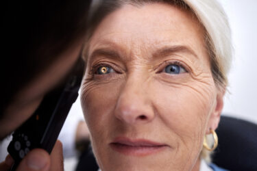
In my opinion, 4 exams constitute a minimum standard of care for monitoring and taking care of glaucoma patients. The first is to look at the optic nerve. This can easily be done with a dilated eye exam, using a lens. It is important that eye care providers become comfortable looking at optic nerves, making sure they have a good view and can evaluate what they see.
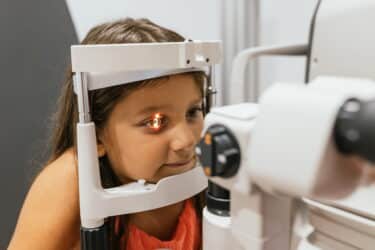
Having some type of optical coherence tomography to look at the retinal nerve fiber layer and ganglion cells is also important, as is measuring eye pressure because that is what will ultimately be treated. Finally, it is important to test the visual field to look for loss of peripheral vision. For our patients, maintaining peripheral vision is their number 1 goal.
I work in a tertiary referral center, where we do a lot of glaucoma evaluation and treatment, and when I carry out a glaucoma evaluation, I offer additional diagnostics. I will definitely measure corneal thickness. I consider pachymetry very important because an eye pressure value measured with a device that does not take into account how thick the cornea is can mean many different things, depending on how thick that patient’s cornea is and how well that patient’s cornea can absorb shock. For this reason, we also like to evaluate corneal biomechanics by measuring corneal hysteresis.
Gonioscopy is a valuable tool because it identifies whether a patient has angle closure glaucoma or open angle glaucoma. These conditions are treated very differently.
It is also nice to have optic nerve head photos. Optic nerve photos allow a visual comparison of nerve cupping which can be easily referenced year after year.
Benefitting from Advancements
There are several exciting new technologies in glaucoma. First is a technology available now, the FAT1 (Falck). This is an attachment device for measuring intraocular pressure as well performing ophthalmodynamometry and tonography. Ophthalmoldynamometry is a surrogate calculation that can be used for ocular perfusion pressure. Tonography looks at the rate of aqueous outflow. This information can help tailor the approach for medical or surgical glaucoma treatment.
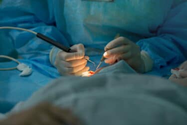
Aqueous angiography is another exciting development on the horizon that will have a sweet spot in surgical glaucoma cases. A better understanding of the anatomy and physiology of a patient’s outflow system will allow surgeons to take a more targeted minimally invasive glaucoma surgery or incisional approach.
Another interesting development, which is still the subject of mixed opinions, is pattern electroretinography. The process allows you to look at the health of the ganglion cells, and pattern electroretinography is mostly positioned as diagnosing glaucoma before there is any change identifiable through optical coherence tomography. Some damage may still be reversible at that point.2
There has also been extensive progress in visual field testing. Automated visual field testing has been in use for roughly 35 to 40 years, and a consistent focus during this time has been how to create faster, reliable tests, that patients can take in a more comfortable, convenient way.
There are new products on the market such as re:ViveTM (Heru) and VisuALL S (Olleyes) that meet these criteria using augmented reality/virtual reality headsets. Re:Vive not only performs full threshold field tests, but also eliminates the need for several legacy devices by performing contrast sensitivity testing, Ishihara and Farnsworth D-15 color vision testing, and dark adaptation testing on commercially available headsets. The platform has shown strong correlation to the gold standard Humphrey Field Analyzer (HFA)3 and Heru has made the platform’s reports and printouts comparable to those produced by the legacy device, so practitioners do not have to learn a new way of reviewing a visual field report.
Increasing Access
These wearable diagnostic platforms can be wall-mounted, so they do not take up valuable floor space in a practice. Our practice is large, and we have 3 HFA’s. We would have a hard time increasing the percentage of patients who receive screening tests on these devices because we would need to move them in and out of the designated testing rooms, and the testing would create a bottleneck. Headsets for visual field testing contain their own lighting environment, so this technology allows testing to be performed comfortably anywhere in the practice. Patients can complete screening between the time the technician leaves the room and the doctor enters or while they are in the waiting room, which makes increased testing more realistic.
For a solo practicing optometrist who does not currently have a visual field device, implementing technology like the re:Vive platform in clinics is a powerful addition in terms of being able to take care of glaucoma patients. If eyecare professionals practice in multiple locations, they can take the lightweight platform with them, and they do not need to add new technology in every practice.
The platform also features a virtual guide that leads the patient through the testing process and monitors fixation in an active way, taking the decision to stop and restart the test out of the technician’s hands. For a large practice like ours, this is great, because it frees up technicians to do other important tasks. For a solo practice with only one technician, the impact of making it possible for that technician to spend more time on activities vital to the practice is even more significant.
With technology that makes screening faster and easier, it may be possible to do visual field testing in response to indicators that currently do not always trigger screenings, such as a family history of glaucoma. Catching glaucoma early is so important and having improved tools for early diagnosis will make a difference for eyecare professionals, their patients, and our healthcare system.
References
- Friedman DS, Wolfs RC, O’Colmain BJ, et al. Prevalence of open-angle glaucoma among adults in the United States [published correction appears in Arch Ophthalmol. 2011 Sep;129(9):1224]. Arch Ophthalmol. 2004;122(4):532-538. doi:10.1001/archopht.122.4.532
- Ventura LM, Porciatti V. Restoration of retinal ganglion cell function in early glaucoma after intraocular pressure reduction: a pilot study. Ophthalmol. 2005;112:20-27.
- Rethinking Glaucoma Management Using Wearable Diagnostics From Heru: re:Vive Visual Field. Seeheru.com. www.seeheru.com/rethinking-glaucoma-management-using-wearable-diagnostics-from-heru-revive-visual-field. Published August 1, 2021. Accessed January 15, 2022.
Mitch Ibach, OD, FAAO
Dr Mitch Ibach is a residency trained optometrist at Vance Thompson Vision in Sioux Falls, SD. He maintains a large collaborative glaucoma clinic that focuses on long-term disease care.
Related Content