Optometry360 Logo White
  • Conferences
  • Videos
  • Podcasts
  • Quizzes
  • About
    • About Us – Mission
    • Content Awards
    • Media Partners
    • Business Team
    • Brand Ambassadors
    • Industry Council
    • Advisory Board

What are you looking for?

  • Anterior Segment
  • Cataract
  • Cornea and External Disease
  • Diabetic Macular Edema
  • Dry Eye
  • Early Onset Cataracts
  • Exclusives
  • General
  • Geographic Atrophy
  • Glaucoma
  • Industry News
  • Inherited Retinal Disease
  • IOLs
  • Neurotrophic Keratitis
  • Ocular Surface Disease
  • Oculoplastics
  • Optometry
  • Pediatrics
  • Practice Management
  • Presbyopia
  • Refractive Surgery/Vision Correction
  • Residents & Young Ophthalmologists
  • Retina
  • Retina Care 360
  • Retinopathy of Prematurity
  • Spotlight Series
  • The Interventional Glaucoma Project
  • The Ophthalmic Project
  • Trending Topics
Spotlight - The Future of Cryopreserved Amniotic Membrane in Oculoplastic Surgery
Optometry360 Logo White
  • Conferences
  • Videos
  • Podcasts
  • Quizzes
  • About
    • About Us – Mission
    • Content Awards
    • Media Partners
    • Business Team
    • Brand Ambassadors
    • Industry Council
    • Advisory Board
Home > Glaucoma > Real-World Data Shows Visual Field Loss Occurs Faster in Glaucoma Patients With Uveitis
  • Glaucoma

Real-World Data Shows Visual Field Loss Occurs Faster in Glaucoma Patients With Uveitis

Ophthalmology 360
Real-World Data Shows Visual Field Loss Occurs Faster in Glaucoma Patients With Uveitis

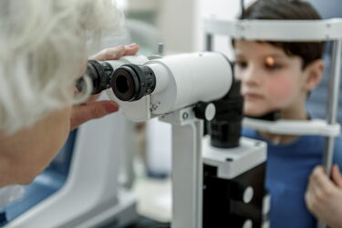
Due to the widespread use of electronic medical records (EMRs), it is now possible to easily collect information from large-scale patient populations to better understand disease progression and treatment response. A new study in the American Journal of Ophthalmology is the first to use this big data approach to compare the rate of visual field (VF) loss in glaucoma patients with uveitis versus those with primary open-angle glaucoma. The study also examined whether the findings were associated with intraocular pressure (IOP).

Anonymous VF and IOP measurements were retrieved from an EMR in five different glaucoma clinic regions in England and included 205 eyes with uveitis plus glaucoma and 4,600 eyes with primary open-angle glaucoma only. Only one eye per patient was analyzed. Minimum inclusion criteria were four or more visits in 4 years. Median patient age was 64 years in the uveitis group and 70 years in the primary open-angle glaucoma group. Researchers used a mixed-effects model and pointwise VF progression analysis of pattern deviation to confirm differences between groups.

Regarding rate of VF loss, the uveitis group had 23 (11%) eyes and the primary open-angle glaucoma group had 331 (7%) eyes that progressed at ≥1.5 dB/year. The relative risk of a fast rate of progression for uveitis was 1.6 and age-adjusted was 1.9, meaning a patient in the uveitis group was 1.9 times more likely to be a fast progressor than patients in the primary open-angle glaucoma group.

Regarding IOP, a total of 143 eyes from the uveitis group and 3,386 eyes from the primary open-angle glaucoma group met the additional inclusion criteria for longitudinal IOP analysis. No statistically significant difference was found in mean IOP between groups. Mean difference in IOP between fast and non-fast progressors was within 1 mmHg.

Results showed that glaucoma patients with uveitis were more likely to be younger and have worse mean deviation at baseline than those with primary open-angle glaucoma. This finding is important because it suggests early VF loss in patients with uveitis may be undetected. However, despite patients with uveitis having a more rapid rate of vision loss, both groups monitored with VFs at the same intensity.

Patients with uveitis require closer attention for ruling out VF loss during treatment.

Reference:

Rabiolo A, Caprioli J. Cataract surgery and rate of visual field progression in primary open-angle glaucoma [published online ahead of print June 2019]. Am J Ophthalmol. doi: 10.1016/j.ajo.2019.04.028.

Share

Related Content

  • Glaucoma

Geographic differences in glaucoma care raise concerns about equitable access

  • Glaucoma

Study finds significant IOP reductions with iDose TR and MIGS procedures

  • Glaucoma

EyeValve device lowers IOP and maintains control for 9 months in first-in-human study

  • Glaucoma

GLP-1 therapies may offer potential benefits in glaucoma

  • Glaucoma

PreserFlo MicroShunt shows short-term benefit in refractory childhood glaucoma

  • Glaucoma

iStent adoption rises as surgeons treat glaucoma earlier

Share

Editor's Picks

  • Neurotrophic Keratitis

Topical insulin shows real-world benefit in neurotrophic keratopathy

  • Retina

GLP-1 RAs have protective effects against AMD

  • Retina

Four-month injection intervals appear safe for long-term stable nAMD

Advisory Board

Saad Ahmad, MD

Ahmad A. Aref, MD, MBA

Roomasa Channa, MD

David Chow, MD, FRCS(C)

Sally L. Baxter, MD, MSc

Neel R. Desai, MD

Nadia Haqqie, MD

Simon Fung, MD, FRCOphth

Sumit Garg, MD

Ross Lakhanpal, MD, FACS

Sanjai Jalaj, MD

Anton Kolomeyer, MD, PhD

Shan Lin, MD

Steven R. Sarkisian, Jr., MD

See All
Optometry360 Logo

Ophthalmology 360® is a dynamic digital platform dedicated to advancing the field of eye care.

Get to Know Us

  • Home
  • About Us
  • Media Partners
  • Advertising Policy
  • Our Advisory Board

Sign up for our Newsletter

Sign up for our Newsletter to get our newest articles instantly!

  • Privacy Policy
  • Advertising Policy
  • Medical Disclaimer
IHM Logo

2026 Ophthalmology 360 is a trademark of International Healthcare Media, LLC. All rights reserved

  • MedJournal360 Icon
  • RareDisease360 Icon
  • MyHero360 Icon
  • Optometry360 Icon
  • Ophtalmology360 Icon