Corneal Edema Following Intraocular Surgery: Current Thinking, Future Directions


By Robert J. Weinstock, MD
Summary:
- Most patients undergoing intraocular surgery will experience some degree of corneal edema
- In most cases, postoperative corneal edema resolves, but sustained corneal edema can have severe vision-threatening consequences
- Risk factors for sustained corneal edema include pre- existing corneal endothelial dystrophies, glaucoma, diabetes, chronic uveitis, and surgical trauma
- Care to protect the corneal endothelium during surgery (e.g., with OVDs) can help minimize risk of corneal edema or endothelial cell loss
- Short-term edema may be treated with anti-inflammatories and topical hypertonic saline
- Longstanding edema may require surgical intervention, which is challenged by the slow recovery of the corneal endothelium
- Future approaches to aid cell protection and proliferation may include the engineered fibroblast growth factor TTHX1114
Introduction
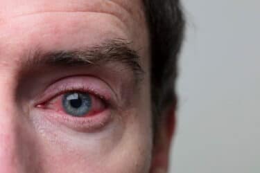
Almost all patients who undergo intraocular surgery will have some degree of corneal edema postoperatively, which can range from superficial epithelial swelling to full-thickness edema.1 In the setting of a normally functioning endothelium, acute corneal edema usually resolves completely, but in some cases, it may be sustained. Vision-threatening sequelae of persistent corneal edema can include limbal stem cell dysfunction, corneal vascularization, surface irregularity, scarring, and even posterior segment changes, such as cystoid macular edema.2 Bullous keratopathy may develop if corneal endothelial cells are damaged irreversibly.3
Candidates for intraocular surgery may present with a pre-existing low endothelial cell count, which puts the cornea at risk of decompensation even after uncomplicated surgery.2 In addition to preoperative endothelial cell density (ECD), the amount of endothelial cell loss that occurs following procedures, such as cataract or minimally invasive glaucoma surgery (MIGS), depends on several risk factors.4
Preoperative risk factors include conditions such as pre-existing corneal endothelial dystrophies, glaucoma, and chronic uveitis.5 Results from a very large-scale analysis6 of 34,234 eyes indicated that factors significantly associated with lower ECD included female sex, older age, Hispanic ethnicity, diabetes mellitus, glaucoma, and a history of refractive or cataract surgery (all P <0.05). It has also been shown7 that dry eye disease is associated with lower ECD (P = 0.046). Research suggests a linear relationship between the severity of pre-existing disease and the degree of postoperative corneal edema and long-term ECD loss.8,9
Intraoperatively, variables, such as instrument trauma, duration of surgery, and vitreous loss, can influence the risk of postoperative edema.
Finally, increased intraocular pressure (IOP), vitreous prolapse, and chronic inflammation in the postoperative period may also put patients at risk of corneal edema and endothelial cell loss.5
Mechanisms
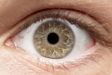
The corneal endothelium is a single layer of cubic cells with high metabolic activity. Its main function is to maintain the translucency of the cornea by preventing the entry of fluid from the anterior chamber. An active ion pump system and the carbonic anhydrase enzyme move ions and water from the corneal stroma into the anterior chamber.4 Corneas with insufficient ECD cannot pump fluid efficiently, which can lead to edema, opacity, and loss of vision.10
Unfortunately, endothelial cells cannot divide and proliferate, and their numbers decrease with age.4 Humans are born with an average corneal ECD of approximately 3000 cells/mm,2 which decreases by adulthood to about 2,500 cells/mm.2,11 As ECD decreases over time, the risk of corneal edema increases.4 Intraocular surgery has the potential to reduce these numbers even further. Even in the absence of complications, surgery may reduce ECD by 9% over 12 months following the procedure.5,12
In comparison, in a healthy individual, endothelial cells decrease by roughly 0.6% per year.4 Depending on the surgical approach, endothelial cell loss resulting from intraocular surgery may increase up to 40% when compared with the healthy fellow eye over an average follow-up of 11 years.13,14 Oxidative stress has been associated with the development of various ocular diseases, such as age-related macular degeneration, cataract, and glaucoma, as well as being a hallmark of Fuchs endothelial corneal dystrophy (FECD).
Oxidative stress occurs when there is an oxidant-antioxidant imbalance due to either the depletion of antioxidants, excess accumulation of reactive oxygen species, or both. This imbalance causes cells to restore balance by activating or silencing genes responsible for functions such as transcription factors, defensive enzymes, and apoptotic pathways. The corneal endothelium is especially prone to oxidative damage due to exposure to light, high oxygen demand, and metabolic activity, as well as the non-proliferative nature of the cells.15
Cataract Surgery
While cataract surgery has been proven to significantly restore vision and productivity in more and more patients, virtually all patients experience corneal edema in the early postoperative period following routine cataract surgery.1,16,17 Depending on the severity of corneal edema and associated inflammation, patients may experience pain, photophobia, or limited vision during the initial period following the procedure.5 Estimates vary, but as many as 1% of cataract surgery patients experience sustained corneal edema, also known as pseudophakic bullous keratopathy.18 Otherwise successful cataract surgery may be jeopardized when the cornea, which accounts for at least two-thirds of the eye’s total refractive power, is compromised as a result of the surgical procedure.19,20
The degree of edema is typically related to the density of the cataract, the surgical time, and the amount of phacoemulsification energy used. As a rule, eyes with denser cataracts tend to have more corneal edema on postoperative day one.4 Following surgery, the most substantial ECD loss occurs in the first month, but the rate of ECD loss continues to be above the normal physiological level for years postoperatively when compared to healthy, unoperated eyes.4,21
In cataract surgery, corneal endothelial damage may occur as a result of ultrasonic energy, fluidics, and surgical trauma, which can stress the cells’ ion pump mechanisms. As a result, corneal clarity is not restored until the endothelial cells recover from the trauma.5 Mild edema often clears within a day or two, but patients who have extremely dense cataracts, whose endothelium is compromised, and/or who have FECD, can have corneal edema that may persist even beyond 6 weeks.2
Sustained corneal edema following cataract surgery, while occurring in a minority of patients, can have a significant and negative impact on vision quality. Long-standing corneal edema may be associated with bullae, which can rupture and lead to severe photophobia and pain.5
Glaucoma Surgery
Reduced ECD in eyes with glaucoma is largely attributed to elevated IOP and is affected by factors, such as disease type, duration, progression, and stability.4 Chronic use of topical anti-glaucoma medications can also have a detrimental effect on the health of the cornea due to the toxicity of preservatives. These variables can affect a glaucoma patient’s baseline ECD and increase the likelihood that they will experience corneal edema following MIGS or other glaucoma surgery.4
The extent of the effect of glaucoma surgery on the corneal endothelial status is strongly dependent on the complexity of the procedure. In general, the more advanced a patient’s glaucoma, the worse their baseline corneal endothelial status, and the more invasive the necessary surgery may have to be.4 Depending on the glaucoma procedure performed, mean ECD loss can be up to 18.6% over 2 postoperative years.4 Results from a meta-analysis of 39 studies revealed that mean ECD loss after glaucoma surgery could be up to 338 cells/mm2 after 12 months.22
Often, MIGS procedures are combined with cataract surgery.23,24 Results from several studies indicate that combining procedures results in equivalent or smaller reductions in ECD compared to performing each procedure separately.25,26 When combined with cataract surgery, MIGS has mostly been shown to be safe in terms of endothelial cell loss.27,28 Notably, however, the CyPass microstent was withdrawn from the market in 2018 due to adverse effects on patients’ corneal endothelial status. Patients who had combined phacoemulsification and CyPass implantation experienced a corneal endothelial cell loss of 20.5% at 5 years, compared to 10.1% in patients who had phacoemulsification alone.4
Intraocular Refractive Surgeries
Intraocular refractive surgeries are often considered for patients with dry eye disease, thin corneas, or extreme myopia or hyperopia that would make them poor candidates for corneal refractive procedures.29
Corneal edema and endothelial cell loss occur in around 10% of eyes implanted with phakic intraocular lenses (pIOLs) over a 5-year period.30 Older age, longer axial length, and smaller anterior chamber depth are risk factors associated with corneal edema in the context of iris-fixated pIOLs.31
Preventing and Treating Postoperative Corneal Edema
In cataract, MIGS, and pIOL surgeries, use of ophthalmic viscoelastic devices (OVDs) help protect the corneal endothelium and are useful especially in patients who are at high risk of corneal edema or endothelial cell loss, such as those with very dense cataract or pre-existing corneal dystrophy. Ophthalmic viscoelastic devices can help protect the cornea from intraoperative damage and ensure a faster recovery.32
Treatment of mild postoperative corneal edema can be accomplished with standard topical anti-inflammatories, including corticosteroids and non-steroidal anti-inflammatory drugs (NSAIDs). For patients with significant corneal edema, the standard of care is topical hypertonic saline. This helps to clear the cornea and draws some of the fluid out, helping to improve the function and recovery of the endothelium.2
If corneal edema is persistent for longer than 2 or 3 months, however, it is unlikely to resolve without surgical intervention, typically a partial thickness transplant, such as a Descemet’s membrane endothelial keratoplasty (DMEK) or a Descemet’s stripping endothelial keratoplasty (DSEK) technique.2
Descemetorhexis without endothelial keratoplasty (DWEK) is an experimental procedure for treating sustained corneal edema where endothelial cells from the periphery migrate and cover the bare stroma.33 This technique is more recently being described as Descemet stripping only (DSO). Due to the protracted time for visual recovery associated with the slow proliferation of endothelial cells, this novel approach can be combined with a rho kinase inhibitor such as ripasudil or netarsudil.33,34
Patients with FECD have a high risk of postoperative corneal edema and of needing a secondary corneal procedure. For these patients, because of the risk of long-term corneal compromise, it makes sense to avoid placing a multifocal IOL.35
Future Prophylactic Approaches
Despite advances in surgical technique and instrumentation and the prophylactic use of OVDs, corneal edema and endothelial cell loss following intraocular surgery remain problems in need of a more reliable preventive solution.
Trefoil Therapeutics’ engineered fibroblast growth factor (FGF), TTHX1114, is under investigation as an intracameral injection for its ability to protect and regenerate corneal endothelial cells.10,35,36 Native FGF1 is a potent stimulator of cell proliferation and migration but is unsuited to pharmaceutical use because of its short half-life. By introducing mutations to FGF1, TTHX1114 is designed to provide a longer half-life with the same protective and proliferative features of endogenous FGF-1.35,36
The safety and efficacy of TTHX1114 are being evaluated in a Phase 2, open-label, dose-ranging clinical trial for patients with corneal endothelial dystrophy who undergo DSO surgery.37
Should fibroblast growth factor prove effective in humans with corneal endothelial dystrophies, it may also prove effective for preventive use in patients undergoing intraocular surgery who are at risk of corneal complications.
A topical formulation of TTXH1114 is also in preclinical development for use in the management of several conditions that lead to corneal ulcers, and should enter clinical testing later this year.38
____
Robert J. Weinstock, MD, is Director of Cataract and Refractive Surgery at the Eye Institute of West Florida, in Largo, FL.
Disclosures: Consultant/Advisor, Lecture fees/Speakers bureau: Bausch & Lomb, Alcon, J&J Vision; Consultant/Advisor, Grant Support: LENSAR; Consultant: Beyeonics, BVI. Equity/Stock holder, public corporation: EyeSafe, RPS, Visus, ViaLase, UBeam, TrueVision, Tissuetech, RxSight, Alchemy Vision Project
References
- Fabrykowski M, Garston M. Comanaging cataract surgery complications. Rev Optometry. 2016; 36-42.
- Srinivas S, Mohanan A, Rao RK. Ten pearls for the management of pseudophakic corneal edema. Ophtha. 2021. https://www.eophtha.com/posts/ten-pearls-for-the-management-of-pseudophakic-corneal-edema.
- Yi DH, Dana MR. Corneal edema after cataract surgery: incidence and etiology. Semin Ophthalmol. 2002;17(3-4):110-114.
- Obuchowska I, Konopinska J. Corneal endothelial cell loss in patients after minimally invasive glaucoma surgery: current perspectives. Clin Ophthalmol. 2022;16:1589-1600.
- Sharma N, Singhal D, Nair SP, et al. Corneal edema after phacoemulsification. Indian J Ophthalmol. 2017;65(12):1381-1389.
- Kwon JW, Cho KJ, Kim HK, et al. Analyses of factors affecting endothelial cell density in an eye bank corneal donor database. Cornea. 2016;35(9):1206-1210.
- Kheirkhah A, Saboo US, Abud TB, et al. Reduced corneal endothelial cell density in patients with dry eye disease. Am J Ophthalmol. 2015;159(6):1022-1026.
- Reinprayoon U, Jermjutitham M, Kasetsuwa N. Rate of cornea endothelial cell loss and biomechanical properties in fuchs’ endothelial corneal dystrophy. Front Med (Lausanne). 2021;8:757959.
- Lundberg B, Jonsson M, Behndig A. Postoperative corneal swelling correlates strongly to corneal endothelial cell loss after phacoemulsification cataract surgery. Am J Ophthalmol. 2005;139(6):1035-41.
- Pizzuto S, Duffey G, Weant J, et al. Acceleration of regeneration of the corneal endothelial layer after Descemet stripping induced by the engineered FGF TTHX1114 in human corneas in organ culture. Cornea. 2022.
- Vaiciuliene R, Rylskyte N, Baguzyte G, et al. Risk factors for fluctuations in corneal endothelial cell density (Review). Exp Ther Med. 2022;23(2):129.
- Bourne RR, Minassian DC, Dart JK, et al. Effect of cataract surgery on the corneal endothelium: modern phacoemulsification compared with extracapsular cataract surgery. Ophthalmology. 2004;111(4):679-685.
- Yeniad B, Corum I, Ozgun C. The effects of blunt trauma and cataract surgery on corneal endothelial cell density. Middle East Afr J Ophthalmol. 2010;17(4):354-8.
- Sminia ML, Odenthal MT, Wenniger-Prick LJ, et al. Traumatic pediatric cataract: a decade of follow-up after artisan aphakia intraocular lens implantation. J AAPOS. 2007;11(6):555-558.
- Ong Tone S, Kocaba V, Böhm M, et al. Fuchs endothelial corneal dystrophy: the vicious cycle of fuchs pathogenesis. Prog Retin Eye Res. 2021;80:100863.
- Blancafort Alias S, Del Campo Carrasco Z, Salvador-Miras I, et al. Exploring vision-related quality of life: a qualitative study comparing patients’ experience of cataract surgery with a standard monofocal IOL and an enhanced monofocal IOL. Clin Ophthalmol. 2022;16:1641-1652.
- Bamdad S, Ahmad Razavizadegan S, Farvardin M, et al. Vision-related quality of life after bilateral implantation of monofocal and multifocal intraocular lenses. J Ophthalmic Vis Res. 2022;17(1):19-26.
- Pricopie S, Istrate S, Voinea L, et al. Pseudophakic bullous keratopathy. Rom J Ophthalmol. 2017;61(2):90-94.
- Shaw E, Patel BC. Complicated cataract. National Library of Medicine. Updated July 5, 2022. Accessed September 19, 2022. https://www.ncbi.nlm.nih.gov/books/NBK572139/ 20. Palanker D. Optical properties of the eye. American Academy of Ophthalmology. Updated October 28, 2013. Accessed September 23, 2022. https://www.aao.org/munnerlyn-laser-surgery-center/optical-properties-of-eye
- Palanker D. Optical properties of the eye. American Academy of Ophthalmology. Updated October 28, 2013. Accessed September 23, 2022. https://www.aao.org/munnerlyn-laser-surgery-center/optical-properties-of-eye
- Bourne WM, Nelson LR, Hodge DO. Continued endothelial cell loss ten years after lens implantation. Ophthalmology. 1994;101(6):1014-22; discussion 1022-3.
- Fang CEH, Mathew RG, Khaw PT, et al. Corneal endothelial cell density loss after glaucoma surgery alone or in combination with cataract surgery: a systematic review and meta-analysis. Ophthalmology. 2022;129(8):841-855.
- Riesen M, Funk J, Töteberg-Harms M. Long-term treatment success and safety of combined phacoemulsification plus excimer laser trabeculostomy: an 8-year follow-up study. Graefes Arch Clin Exp Ophthalmol. 2022;10.1007/s00417-021- 05510-8.
- Moreno-Valladares A, Puerto Amorós N, Mendez Llatas M, et al. Combined excimer laser trabeculostomy and phacoemulsification: one year follow-up real world data of a laser-based MIGS. Arch Soc Esp Oftalmol (Engl Ed). 2021;96(12):631-639.
- Soro-Martínez MI, Miralles de Imperial-Ollero JA, Pastor-Montoro M, et al. Corneal endothelial cell loss after trabeculectomy and phacoemulsification in one or two steps: a prospective study. Eye (Lond). 2021;35(11):2999-3006.
- Demir AG, Olgun A, Guven D, et al. The effect of combined phacotrabeculectomy, trabeculectomy and phacoemulsification on the corneal endothelium in the early stage: a preliminary study. Int Ophthalmol. 2019;39(9):2121-2128.
- Vold S, Ahmed II, Craven ER, et al. Two-year COMPASS trial results: supraciliary microstenting with phacoemulsification in patients with open-angle glaucoma and cataracts. Ophthalmology. 2016;123(10):2103- 2112.
- Ahmed IIK, Rhee DJ, Jones J, et al. Three-year findings of the HORIZON trial: a Schlemm canal microstent for pressure reduction in primary open-angle glaucoma and cataract. Ophthalmology. 2021;128(6):857-865.
- Kozak A, Buckner B, Reddy V, et al. American Academy of Ophthalmology EyeWiki. Updated April 10, 2022. Accessed September 6, 2022. https://eyewiki.aao.org/Intraocular_Refractive_Surgery
- Jonker SMR, Berendschot TTJM, Ronden AE, et al. Five-year endothelial cell loss after implantation with artiflex myopia and artiflex toric phakic intraocular lenses. Am J Ophthalmol. 2018;194:110-119.
- Jonker SMR, Van Averbeke AAC, Berendschot TTJM, et al. Risk factors for explantation of iris-fixated phakic intraocular lenses. J Cataract Refract Surg. 2019;45(8):1092-1098.
- Borkenstein AF, Borkenstein EM, Malyugin B. Ophthalmic viscosurgical devices (OVDs) in challenging cases: a review. Ophthalmol Ther. 2021;10(4):831-843.
- Rodríguez-Fernández S, Piñeiro-Ramil M, Castro-Viñuelas R, et al. Current development of alternative treatments for endothelial decompensation: cell-based therapy. Exp Eye Res. 2021;207:108560.
- Syed ZA, Rapuano CJ. Rho kinase (ROCK) inhibitors in the management of corneal endothelial disease. Curr Opin Ophthalmol. 2021;32(3):268-274.
- Kaup S, Pandey SK. Cataract surgery in patients with fuchs’ endothelial corneal dystrophy. Community Eye Health. 2019;31(104):86-87.
- Eveleth D, Pizzuto S, Weant J, et al. Proliferation of human corneal endothelia in organ culture stimulated by wounding and the engineered human fibroblast growth factor 1 derivative TTHX1114. J Ocul Pharmacol Ther. 2020;36(9):686-696.
- Pizzuto S, Weant J, Afshari NA, et al. Accelerated regeneration of corneal endothelium after Descemet’s stripping mediated by the engineered fibroblast growth factor TTHX1114. Invest Ophthalmol Vis Sci. 2021;62(8):797.
- A safety and efficacy trial of TTHX1114 in people with CED (OPTIC). ClinicalTrials. gov identifier: NCT04812067. https://clinicaltrials.gov/ct2/show/NCT04812067. Updated January 25, 2022. Accessed September 8, 2022.
Related Content