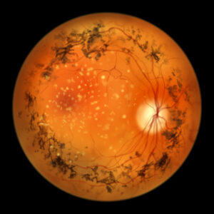
New imaging technique provides insights into retinitis pigmentosa progression

A recent study explored the relationship between the structure and function of surviving photoreceptors in patients with retinitis pigmentosa (RP). Using advanced imaging methods, including adaptive optics scanning laser ophthalmoscopy and adaptive optics optical coherence tomography, researchers sought to understand how surviving photoreceptors continue to function despite degeneration.
Key Findings:
- Cone density was significantly reduced in patients with RP, particularly in areas transitioning from healthy to diseased retina.
- Despite reduced cone density, visual sensitivity in 3 out of 4 patients remained comparable to healthy controls.
- The study revealed that optoretinograms (ORGs), which measure functional responses in cones, were diminished in patients with RP, even in regions where cone density appeared normal.
- ORG responses and cone outer segment lengths were correlated in healthy controls but not in patients with RP.
The findings suggest that ORG-based measures can detect retinal dysfunction before visible structural damage or vision loss occurs.
Reference
Wendel BJ, Pandiyan VP, Liu T, et al. Multimodal High-Resolution Imaging in Retinitis Pigmentosa: A Comparison Between Optoretinography, Cone Density, and Visual Sensitivity. Invest Ophthalmol Vis Sci. 2024;65(10):45. doi: 10.1167/iovs.65.10.45. PMID: 39207297; PMCID: PMC11364184.
Related Content