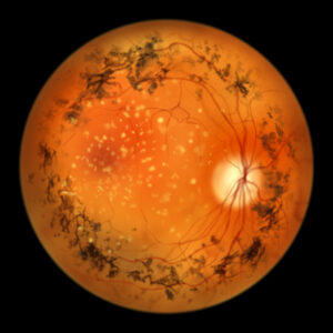
Outer retinal degeneration noted in patients with Stargardt disease over 2-year period

Spectral-domain optical coherence tomography (SD-OCT) has successfully detected significant degeneration in outer retinal layers in patients with Stargardt disease type 1 (STGD1) over a 24-month period, according to a study.
SD-OCT images from 428 eyes of 236 patients were analyzed. The researchers focused on changes in mean thickness and intact area after semi-automated segmentation of various retinal layers in 3 zones of the ETDRS grid: central subfield, inner ring, and outer ring. The layers studied included the retinal pigment epithelium, outer segments, inner segments, outer nuclear layer, inner retina, and total retina.
Over the 24-month period, the study observed significant decreases in all outer retinal layers, including the retinal pigment epithelium, outer segments, inner segments, and outer nuclear layer. This decline was evident in both the mean retinal thickness and the intact area (P < 0.0001). In contrast, the inner retina showed an increase in retinal thickness in the central subfield and inner ring, with no significant change observed in the outer ring.
These findings suggest that changes in thickness and intact area of these layers could serve as valuable endpoints for clinical trials aimed at slowing the progression of Stargardt disease.
Reference
Strauss RW, Lang L, Ho A, et al. The Progression of Stargardt Disease (ProgStar) as determined by spectral-domain optical coherence tomography over a 24-month period (ProgStar Report No. 19). Ophthalmic Res. 2024;doi: 10.1159/000540028. Epub ahead of print. PMID: 39004077.
Related Content