Optometry360 Logo White
  • Conferences
  • Videos
  • Podcasts
  • Quizzes
  • About
    • About Us – Mission
    • Content Awards
    • Media Partners
    • Business Team
    • Brand Ambassadors
    • Industry Council
    • Advisory Board

What are you looking for?

  • Anterior Segment
  • Cataract
  • Cornea and External Disease
  • Diabetic Macular Edema
  • Dry Eye
  • Early Onset Cataracts
  • Exclusives
  • General
  • Geographic Atrophy
  • Glaucoma
  • Industry News
  • Inherited Retinal Disease
  • IOLs
  • Neurotrophic Keratitis
  • Ocular Surface Disease
  • Oculoplastics
  • Optometry
  • Pediatrics
  • Practice Management
  • Presbyopia
  • Refractive Surgery/Vision Correction
  • Residents & Young Ophthalmologists
  • Retina
  • Retina Care 360
  • Retinopathy of Prematurity
  • Spotlight Series
  • The Interventional Glaucoma Project
  • The Ophthalmic Project
  • Trending Topics
Spotlight - The Future of Cryopreserved Amniotic Membrane in Oculoplastic Surgery
Optometry360 Logo White
  • Conferences
  • Videos
  • Podcasts
  • Quizzes
  • About
    • About Us – Mission
    • Content Awards
    • Media Partners
    • Business Team
    • Brand Ambassadors
    • Industry Council
    • Advisory Board
Home > Anterior Segment > New algorithm shows promise for early detection of keratoconus in adolescents
  • Anterior Segment

New algorithm shows promise for early detection of keratoconus in adolescents

Ophthalmology 360

A newly developed algorithm for detecting keratoconus in adolescents using optical coherence tomography (OCT) demonstrated high sensitivity and specificity, aligning closely with current gold-standard diagnostic methods, according to a study. The algorithm focuses on identifying alterations in corneal pachymetric and epithelial thickness.

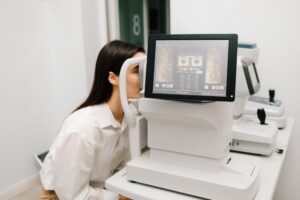
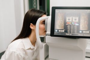
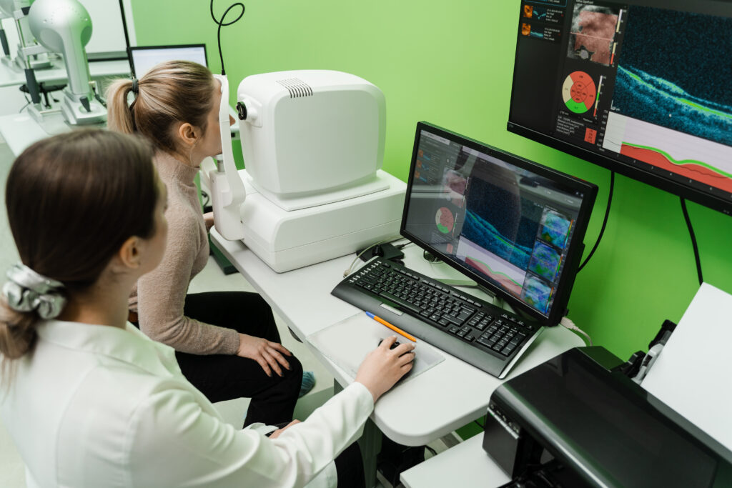
The retrospective study involved reviewing the medical charts of 19 pediatric patients with keratoconus, categorizing them into 4 groups based: normal, manifest keratoconus, subclinical keratoconus, and very asymmetric eye with normal topography and tomography (VAE-NTT). Corneal and epithelial thickness maps were analyzed using the Cirrus 5000 HD-OCT system by a human grader.

The diagnostic process consisted of 2 steps. Initially, the presence of keratoconus was suspected if any of the 4 parameters—pachymetry minimum (pachy min), pachy minimum-median (min-med), pachy superonasal-inferotemporal (SN-IT), or epithelial (epi SN-IT)—exceeded their respective cut-off values. Subsequently, a definitive diagnosis was made if combined thinning of the cornea and epithelium and concentric epithelial thinning were observed.

The algorithm demonstrated impressive diagnostic performance, with area under the curve (AUC) values of 0.889 for pachy min, 0.997 for pachy min-med, 0.893 for pachy SN-IT, and 0.998 for epi SN-IT in the initial step. Overall, step 1 of the algorithm achieved 97.3% sensitivity, while step 2 demonstrated 100% specificity.

This approach, especially when combined with other diagnostic examinations, has the potential to enhance the accuracy of keratoconus detection in adolescents, the authors concluded.

Reference
Yücekul B, Förster A, Dick HB, et al. Detecting Keratoconus in Adolescents with Anterior Segment Optical Coherence Tomography. J Ophthalmol. 2024;2024:6655217. doi: 10.1155/2024/6655217. PMID: 38881564; PMCID: PMC11178420.

Share

Related Content

  • Anterior Segment

AS-OCT analysis shows deeper corneal layer thickening in pseudoexfoliation syndrome

  • Anterior Segment

Study compares tear meniscus measurements in patients with low vitamin D levels

  • Anterior Segment

Global AS-OCT research surges, highlighting growth in AI and imaging innovation

  • Anterior Segment

Age and sex influence key anterior segment metrics in healthy eyes

  • Anterior Segment

Ophthalmologic interventions can improve quality of life in hospice care

  • Anterior Segment

AI’s potential for early diagnosis of vitreoretinal lymphoma

Share

Editor's Picks

  • Neurotrophic Keratitis

Topical insulin shows real-world benefit in neurotrophic keratopathy

  • Retina

GLP-1 RAs have protective effects against AMD

  • Retina

Four-month injection intervals appear safe for long-term stable nAMD

Advisory Board

Saad Ahmad, MD

Ahmad A. Aref, MD, MBA

Roomasa Channa, MD

David Chow, MD, FRCS(C)

Sally L. Baxter, MD, MSc

Neel R. Desai, MD

Nadia Haqqie, MD

Simon Fung, MD, FRCOphth

Sumit Garg, MD

Ross Lakhanpal, MD, FACS

Sanjai Jalaj, MD

Anton Kolomeyer, MD, PhD

Shan Lin, MD

Steven R. Sarkisian, Jr., MD

See All
Optometry360 Logo

Ophthalmology 360® is a dynamic digital platform dedicated to advancing the field of eye care.

Get to Know Us

  • Home
  • About Us
  • Media Partners
  • Advertising Policy
  • Our Advisory Board

Sign up for our Newsletter

Sign up for our Newsletter to get our newest articles instantly!

  • Privacy Policy
  • Advertising Policy
  • Medical Disclaimer
IHM Logo

2026 Ophthalmology 360 is a trademark of International Healthcare Media, LLC. All rights reserved

  • MedJournal360 Icon
  • RareDisease360 Icon
  • MyHero360 Icon
  • Optometry360 Icon
  • Ophtalmology360 Icon