Small aperture optic-based IOL provides an increased range of vision from far to near.
By Elizabeth Yeu, MD
Presbyopia-correcting lens technology continues to expand, providing a growing array of options for our patients. Still, there are limitations when it comes to solutions for patients with irregular corneas and these patients present unique challenges when it comes to postoperative visual results.
In general, there are three groups of irregular cornea patients: (1) surgically-induced such as post-LASIK, PRK, radial keratotomy (RK), astigmatic keratotomy, pterygium surgery, corneal transplants, and trabeculectomy; (2) naturally occurring such as ectasias/keratoconus and corneal dystrophies; and (3) disease or trauma-induced corneal shape irregularities that result in scarring, edema, and other abnormalities.
Research shows that 12% to 14% of patients presenting for cataract surgery have a complex cornea.1 Surgeons participating in a panel discussion on the topic reported that approximately 24% of their preoperative cataract patients have irregular corneas.2 A chart review of 400 eyes in 200 consecutive patients presenting for cataract surgery with no previous corneal surgery revealed that about 25% had abnormal topography.3
Small Aperture, Big Benefits
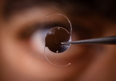
The IC-8 IOL (AcuFocus, Inc), expected to be FDA approved in 2022, gives us the option of leveraging a foundational therapy for irregular cornea patients: pinhole vision. It is well established that pinhole vision or the use of small aperture optics enhances not only depth of vision but quality as well. In an irregular cornea, this potential presbyopia treatment option does three things: provides improved uncorrected distance quality vision; offers an expanded range of vision; and is a non-toric IOL that is indicated for upwards of 1.50 D of irregular astigmatism.4-6
An aspheric monofocal lens with an embedded mask, the IC-8 IOL applies the small aperture principle to extend the depth of focus to provide an increased range of vision from far to near. The wavefront-filtering design eliminates unfocused peripheral light rays so that only the central rays focus on the retina.7 The small aperture design provides an extended depth of focus that makes the implant more tolerant of sphero-cylindrical residual refractive errors compared with multifocal implants.
Studies have shown the IC-8 IOL can provide up to 3.00 D of extended depth of focus and tolerate up to 1.00 D deviation from the target manifest refraction spherical equivalent.8 In contrast, with multifocal technology anything over 0.75 D of residual sphere will compromise visual acuity and spectacle independence, leading to unhappy patients.9 The IC-8 IOL has also been found to enhance vision at all distances, with improved visual acuities and a broad range of continuous functional vision.
The pinhole-based IOL also provides a friendlier landing zone, which is great for the RK patient. I can leave them a little bit myopic, and the pinhole extends their distance and near vision even with the visual fluctuations some of these patients experience throughout the day.
My Experience
The IC-8 IOL is more forgiving when it comes to uncorrected vision. For example, consider a mildly irregular cornea of a keratoconic eye that has great potential for spectacle-corrected vision, to undergo pseudophakic surgical correction with a toric IOL. Getting that patient better near and distance vision postoperatively for any residual astigmatism with a bi-toric rigid gas permeable (RGP) contact lens or other therapeutic lens is a complex process. With the small aperture IC-8 IOL, they will have improved uncorrected, quality distance and near vision. They could still wear an RGP if they needed even more pristine vision, but they will not have to be dependent on it. The small aperture lens is a whole different ballgame, and it is not based on toricity correction through the normal optical manipulation on the back surface of the IOL.
The small aperture effect of the IC-8 can get many presbyopic complex cornea patients out of reading glasses—something we would not have thought possible for this group. In my opinion, the option is superior to some of the extended depth of focus monofocal technologies. Pinhole optics extend the range of uncorrected vision providing quality distance and quality near, while mitigating visual disturbances like glare that comes from the mid-peripheral cornea.
Crucial Topography
One of the most important parts of the workup for all cataract patients, but for these patients in particular, is determining the quality, consistency, and quantity of astigmatism using Placido disc corneal topography. Not all patients know if they have a condition that creates irregular astigmatism or even remember that they have had previous refractive surgery. Surgeons can identify the irregularities topographically and match that up with what is known about their ocular history. The corneal topography can guide me to determine the regularity and irregularities of the cornea, cluing me in to the most appropriate IOL technologies for each individual patient.
Sometimes an over-refraction with a trial RGP can diagnostically assist to discern how much of the visual decline is from the cornea versus the cataract. For example, a patient may have visual acuity of 20/60, but in a rigid gas permeable lens test they see 20/40, therefore 2 lines of their vision is diminished from their cornea. Doing an RGP over-refraction is a fantastic way to figure out if the cornea or the cataract is causing the visual symptoms. When corneal irregularity plays into the patient’s vision problems, correction through small-aperture technology can provide a better overall quality of vision, and provide an extended depth of focus.
Counseling Patients
When counseling irregular cornea patients having cataract surgery, I explain to them a regular, normal cornea provides a clear, pristine path to the retina allowing vision. Whether from trauma, previous surgery, or underlying disease, however, the shape of their cornea is not equal circumferentially, which leads to low-quality rays of light entering the eye at different angles. Without the option of the IC-8 IOL, I would have told previous RK patients that their best-quality vision postoperatively would be achieved by going back to wearing the RGP contact lens. I would caution them that they will still have glare from the RK wounds. When the IC-8 is available, however, we will have an option for them.
Time will tell what best approach to take in terms of IOL calculations is in conjunction with small-aperture technology, but we can look to the international experience for guidance, where the lens has been available for some time. The offsets we currently use such as the post-refractive surgery formulas on the ASCRS calculator and the Barrett True-K formula will be helpful. Within the offset, there is a softer landing zone so even if the target is off by 0.50 D, their uncorrected visual quality will not be compromised.
Conclusion
The IC-8 IOL is implanted in the non-dominant eye, and I see it as having a huge benefit for this segment of the population to be able to achieve an improved quality of uncorrected presbyopia correction. I was very pleased with the results I observed in my experience as a clinical investigator in the FDA trial, and I am hopeful the implant will soon be commercialized.
Elizabeth Yeu, MD, is a cataract, corneal, and refractive surgeon at Virginia Eye Consultants, Norfolk, Virginia. She may be reached at [email protected]. Dr. Yeu was an investigator for the FDA registration study for the IC-8 IOL and is a consultant for AcuFocus.
Reference
- Data on file Acufocus.
- 2020 Global Consensus on Corneal Irregularity: Expert Panel Offers Recommendations for Defining, Diagnosing, and Treating Irregular Corneas. Supplement to Cataract & Refractive Surgery Today / Europe. November/December 2020. https://crstoday.com/wp-content/uploads/sites/4/2021/01/0121CRST-CRSTES_Evolve-2032-Corneal-Irregularity-Consensus-Paper.pdf. Accessed April 29, 2021.
- Frank B, Trattler W, Mccabe S, et al. The incidence of topographic abnormalities in patients scheduled for cataract surgery (abstract). Invest Ophthalmol Vis Sci. 2014; 55:2477.
- Grabner G, Ang RE, Vilupuru S. The small-aperture IC-8 intraocular lens: a new concept for added depth of focus in cataract patients. Am J Ophthalmol. 2015;160(6):1176-1184. doi:10.1016/j. ajo.2015.08.017.
- Dick HB, Piovella M, Vukich J, et al. Prospective multicenter trial of a small-aperture intraocular lens in cataract surgery. J Cataract Refract Surg. 2017;43(7):956-968. doi:10.1016/j.jcrs.2017.04.038.
- Ang RE. Small-aperture intraocular lens tolerance to induced astigmatism. Clin Ophthalmol. 2018;12:1659-1664.
- Tucker J, Charman WN. The depth-of-focus of the human eye for Snellen letters. Am J Optom Physiol Opt. 1975;52(1):3-21. doi:10.1097/00006324-197501000-00002.
- Ang RE. Visual performance of a small-aperture intraocular lens: first comparison of results after contralateral and bilateral implantation. J Refract Surg. 2020;36(1):12-19. doi: 10.3928/1081597X-20191114-01.
- Braga-Mele R, Chang D, Dewey S, et al. Multifocal intraocular lenses: relative indications and contraindications for implantation. J Cataract Refract Surg. 2014;40(2):313–322.
Related Content