During the American Academy of Ophthalmology’s 2018 annual meeting in Chicago, Alan H Zalta, MD, a glaucoma specialist at the Cincinnati Eye Institute, addressed 10 potential pitfalls when using automated threshold perimetry, as well as strategies to overcome these problems.
1. Legally blind eyes. “Many legally blind eyes can provide highly reliable, valuable information about the status of the optic nerve, macular, and retina,” said Dr. Zalta. Be sure to:
- Use nonstandard size V stimulus in the face of dense medial opacity
- Use large diamond fixation when presented with maculopathy
- Monitor fixation visually, and verbally direct it as needed
2. Low reliability message. In these cases, it is likely that the criteria are too rigid, meaning that the reliability message is often incorrect. Graded reliability is more realistic, using “excellent,” “good,” “fair,” and “poor” grades.
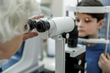
3. Children. “You can perform a reliable field test in children as young as 5 years of age,” he noted. They keys are to:
- Take breaks every few minutes
- Encourage and reinforce the patient
- Optimize alignment and replot or decrease the size blind spot check as needed
- Turn fixation monitor off if necessary
4. Normal gray scale display. Keep in mind that very early glaucomatous defects might be missed. Approximately 2% of visual fields and 10% of eyes with simultaneous “normal” gray scale display scotoma on the pattern deviation plot. “Check the pattern deviation displays before deciding ‘normalcy.’”
5. Normal central 30-degree field and isolated peripheral nasal steps. This occurs in 4% to 11% of eyes with early glaucoma damage. Perform 30/60-2 testing in high-risk glaucoma suspects, including those with severe ocular hypertension, pigment dispersion syndrome, optic disc hemorrhage, and cup-to-disc asymmetry.
6. End-stage visual field loss with intact fixation. Specifically, when visual acuity is ≥20/40, cup-to-disc ratio is >0.9, and central island of vision is <13 degrees. If mean deviation is >-20 decibels (dB), repeat 30-2 testing with size V stimulus. If >-25 dB, perform 10-2 testing with size V stimulus.
7. Glaucoma Hemifield test. Its use to identify early glaucoma defects is limited; there is a 27% false negative rate. Therefore, “always examine pattern deviation displays before [making] any diagnostic or therapeutic decisions.”
8. Miotic pupil artifact. This may cause a pseudo-defect or pseudo-progression. “Dilate all miotic pupils to >2.5 mm for visual field testing. The only exception is [cases of] end-stage glaucoma with intact fixation and small central island of vision in order to avoid idiosyncratic IOP rise.”
9. Ptosis or intermittent droopy upper eyelid artifact. This occurs in 1% to 2% of all central threshold field exams, and may cause pseudo-scotoma or pseudo-progression. “Tape the upper lid to the forehead during field testing, except in eyes with at-risk keratopathy.”
10. Lens rim artifact. Occurring in 9% of all fields in which trial lenses are used, this too may cause pseudo-scotoma or pseudo-progression. To minimize its occurrence:
- Educate the perimetrist and patient
- Use only wire-rimmed, full aperture lenses for central field testing
- Position the head and body to minimize squirming
- Position corrective lenses as close to the globe as possible, but do not touch lashes
- Monitor alignment of the eye and corrective lenses, especially toward the end of testing; reposition when necessary
- Allow short breaks to avoid excessive movement
Reference
Zalta A. Top 10 pitfalls, problem solving, and an interpretive strategy for automated threshold perimetry. Talk presented at: AAO 2018 annual meeting; October, 26-30, 2018; Chicago.
Related Content